The new avatar: from the inside out
When a person moves, you see it on the skin. The shoulder blade glides beneath it, the muscle thickens, the tendon stands out. We want an avatar that can do this, so we are building it the way a body is built: skeleton first, then muscles and fasciae, skin last. Here we report how far we have come, what we have measured and what we do not know yet.
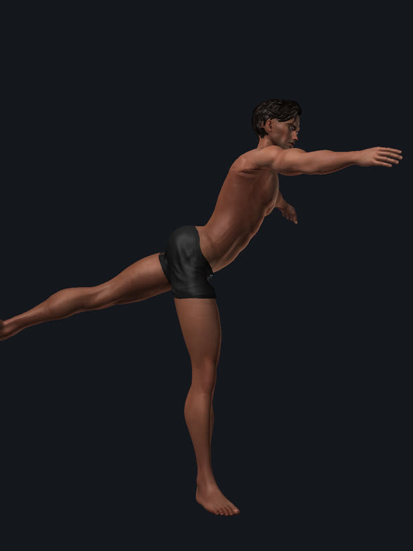
The avatar and the template
The video shows the avatar of a resident of our grid, as the viewer shows it: a commercial mesh body and head in his shape, with his viewer-baked skin, his hair and his swim trunks. New in this version: he no longer moves on the viewer’s Bento skeleton but on our skeleton with 236 bones. A program moved him over automatically. It builds the skeleton in his proportions, every old joint stays exactly where it was, and redistributes the weights of his skin: the trunk onto the 24 vertebrae by height, the foot onto heel bone, metatarsus and every toe, the forearm onto the radius, the shoulder onto collarbone and shoulder blade. At rest the skin then deviates from the old one by less than a micrometre; the program checks this. Underneath lies the anatomy of our template, the open anatomical model BodyParts3D: 934 parts, among them 442 muscle parts and 251 bones, cartilages and discs, computed into his body and likewise moved onto the new skeleton. It remains a single model built from textbooks and atlases. The yardstick for “normal” is therefore measurement studies.
The movements come from classical ballet, because it takes the joints to their limits, and that is where every flaw of a skeleton shows. Every pose is checked against a table of anatomical ranges of motion, with a source per row; the film is only computed if no limit is violated. First position with 50° of outward rotation per hip (measured passively at 54 ± 10° in ballet dancers), demi-plié, relevé: the foot bends at the big-toe joint and stands on the toes, because every toe is a joint; the toes stay flat on the floor, ankle and toe joints at 40° each (recreational dancers lift the heel by 35 ± 4°, Imura 2022; dancers extend the big-toe joint in relevé by 70 to 90°, Veirs 2023). Port de bras through first, second and fifth position, the hand softly grouped, the fingers in the functional posture that Hume 1990 measured during eleven everyday tasks (knuckle 61°, middle joint 60°, end joint 39°), the thumb towards the palm. Tendu and dégagé with a pointed foot, cambré backwards and to the side through all 24 vertebrae. The arabesque follows a measurement of 25 professional dancers (Feipel 2003): the pelvis tips forward over the standing hip, the lumbar spine extends by about 20° as it does, and the working leg rises with the hip extended, the trunk bends 22° towards the working leg and turns 11° away from it, as Feipel measured. The floor is a constraint: when a foot lands it rolls through the ball, ankle first, then the hip, never beyond the limit. Four measurement gates run over every frame: no hand point inside the trunk or thigh, no working foot below the floor, toes flat in relevé, and the fascia nowhere stretches more than the skin. Every movement accelerates and brakes smoothly (Flash and Hogan 1985), distant limbs follow with a small delay, the shoulder blade rotates when the arm is raised (Scibek 2012), above 90° the upper arm turns outward (Inui 2009), pronation is a rotation of the radius. He breathes and sways slightly. Then the peeling away: skin, fascia, muscles, skeleton, joint chain.
What this version does not show: the muscle bulge when shortening and the skin gliding over the muscles, which today’s viewers do not show either. Tendons are computed, not taken from data; the fascia is his own skin offset three millimetres inward, with the template’s relief where it lies close beneath – the template’s fascia spanned a web across the armpit that fanned out when the arm was raised. The height of the arabesque is below that of the professionals. The limits for hip and knee flexion, hip extension, ankle, shoulder elevation, elbow and forearm rotation are means of 143 women from the open CDC reference dataset (Soucie 2011); hip abduction and rotation, shoulder rotation, wrist and the ballet hand still come from teaching texts and physiotherapy tables. Ribs, kneecap and metacarpus carry no weights yet after the transfer.
Anatomical model: BodyParts3D, © The Database Center for Life Science, CC BY-SA 2.1 Japan; rendered by us, the anatomy layers are under the same licence. The avatar in the video belongs to its owner; body and head are purchased third-party products, reproduced as in the viewer; it is not under this licence.
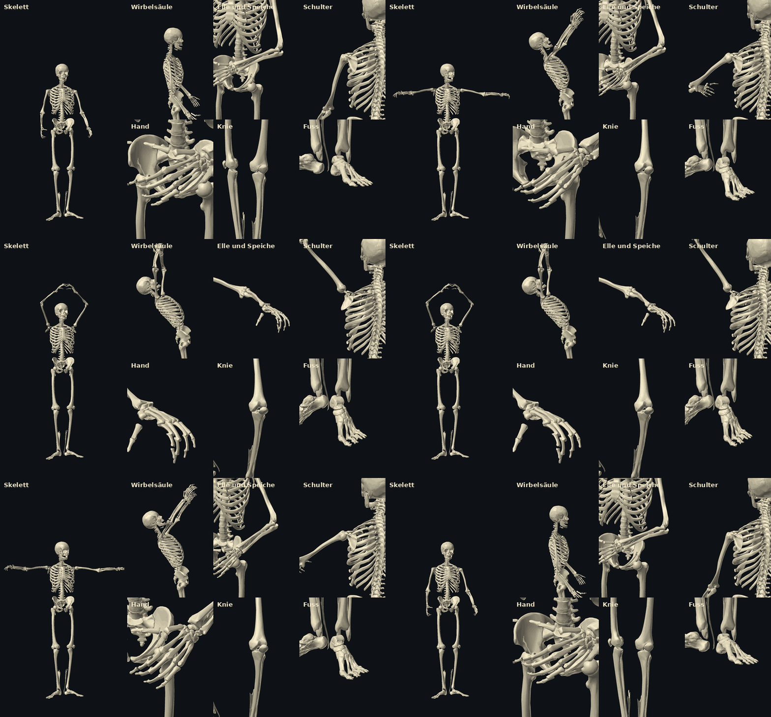
The skeleton
The standard skeleton of today’s viewers has 133 bones, many of them in the face. Our current version has 236. New are 24 separate vertebrae, sacrum and coccyx, a joint between skull and atlas, collarbone and shoulder blade, ulna and radius, the palm, the kneecap, groups for the ribs and a foot with heel bone, midfoot and every toe.
Existing content should keep working. So every previous bone still sits, by calculation, exactly where it was, including across all 71 body sliders. Our programs check this.
The picture shows the bones of the BodyParts3D template, fitted into this avatar’s body and moved on our skeleton, in views from the same ballet as in the video. Kneecap and ribs still follow their neighbouring bones, without motion of their own. The current version does not run in any viewer yet: that needs a viewer build of our own.
A spine in an S-curve
A straight column of separate vertebrae would not yet be a human spine. So our curve comes from a measurement study of 100 adults: lumbar lordosis 56.4°, thoracic kyphosis 44.4°. Much of the lordosis sits right at the bottom, just under 43% between L5 and S1 alone (study of 324 adults). The cervical lordosis of 17.1° comes from a third study.
A mistake belongs in this report: at first we took the wrong table value for the lumbar lordosis. Rereading the table exposed it, and an unexplained kink in the curve disappeared.
One compromise remains: the head has to stay in place for existing content, so our whole curve is tilted by about 7°. The angles between sections follow the studies, but the sacral slope is 33° instead of the measured 40° or so.
What you see on the skin
Skin glides. In a CT study of ten healthy men, skin markers over the lower angle of the shoulder blade travelled about 70 mm when the arm was raised, and reproduced the bone’s movement only partly: 27° instead of 40.5° of upward rotation. Skin fixed rigidly to the bone would be wrong there.
Muscles thicken. In an ultrasound study, the tibialis anterior grew 2.5 mm thicker at half of maximum force, while its volume stayed about the same. Such values hold for this very muscle. For most others we have not yet found reliable measurements.
Fasciae are more than packaging. These connective-tissue sheaths let layers glide against each other and link muscles together. In the lumbar fascia, researchers measured about 70% shear strain by ultrasound in people without back pain and about 56% in people with chronic back pain. We still want data to show how they shape what is visible in places like the palm or the gluteal fold. Our anatomical model contains hardly any fasciae; this layer must come from other sources.
The hardest question: how does it reach the skin?
Viewers of this kind move the skin through bones: each point follows at most four bones, and a mesh can use at most 110 bones (as the Firestorm source code states). That is enough for rotations, but a bulging muscle can probably be shown only roughly. That would take corrective shapes, which the mesh code we checked does not know. To our knowledge no other viewer has shipped them either.
So we will test on a single joint what bones alone achieve and what only corrective shapes bring. The result decides how far we extend our own viewer.
What is still open
- The body in the video is a resident’s avatar, moved automatically onto our 236-bone skeleton, computed for the film, not in a viewer: there is no viewer build of our own yet, and the transferred mesh cannot be tried on in-world yet. His anatomy is computed in from the template, not measured on him.
- The positions of the most recently added bones are estimates. We are replacing them with measurements on the template.
- The standard avatar’s proportions may differ from human ones. So far this is only a suspicion, which we are checking against population data.
- Ribs exist only as groups for now, and the skeleton does not yet enforce movement limits.
- For many joints, muscles and fasciae we still lack open primary data.
The next steps
- Measure bone positions on the template and check proportions against population data.
- Build our own viewer as soon as its source is available.
- Test one joint with skin: bones only against corrective shapes.
- Move existing avatars onto the new skeleton automatically: the converter runs for the avatar in the video; what is missing is the output as a mesh for the viewer.
Sources
- Spine 1: PMC8873993
- Spine 2: PMC9643535
- Spine 3: PMC6942398
- Skin and shoulder blade: PMC9460682
- Muscle thickness: PMC4974924
- Lumbar fascia: PMC3189915
- External rotation during arm elevation: Inui 2009, PMC2823194
- Resting arm position (upright CT): J Orthop Surg Res 2022
- Spiraldynamik, systematics: Larsen, Deswarte
- Velocity profile of arm movements: Flash and Hogan 1985
- Forward bending: lumbar spine and hips: Esola 1996
- Shoulder blade during arm raising: Scibek 2012, PMC3377910
- Lumbar spine in ballet postures, arabesque: Feipel 2003, ISB
- Hip turnout in ballet dancers: PMC12574594
- Functional posture of the finger joints: Hume 1990
- Ankle in demi-pointe: Imura 2022
- Big-toe joint in relevé: Veirs 2023
- Joint range of motion reference values, open dataset: CDC, Soucie 2011
- Overlapping action, moving hold: Lasseter 1987 after Thomas and Johnston 1981
Our summary; not a medical statement. Each study measures specific groups and conditions.